


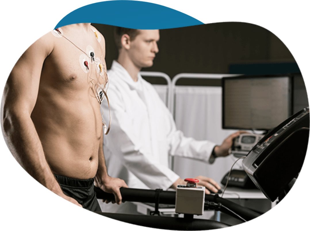
Nuclear stress testing helps medical providers evaluate how well the heart functions under stress. The procedure uses a small amount of radioactive tracer, and a specialized camera captures detailed images of blood flow. These images allow doctors to assess circulation patterns, and they review how the heart responds during activity and at rest.
Understanding Nuclear Stress Testing
Nuclear stress testing provides insight into how blood moves through the heart muscle. The scan produces clear and detailed images. Tissue from a past heart attack may appear on the scan, and medical providers evaluate the extent of this injury. A normal scan indicates healthy heart muscle, and the images show steady blood flow throughout the heart. Because irregular patterns can indicate narrowed arteries, doctors look for specific signs of blockages, and they determine the location of the concern.
Doctors also compare rest and stress images to identify changes in blood flow. This comparison highlights functional differences. When blood flow appears reduced during activity, providers assess the severity of the issue, and they determine whether further evaluation is necessary. Some patients complete exercise-based testing, and others receive medication to simulate stress on the heart. These variations allow providers to gather consistent data, and they ensure the results reflect how the heart performs under different conditions.
Guiding Medical Treatments
Physicians use these scans to support medical decisions. The results inform treatment planning. The test results help guide therapy plans, and doctors adjust medications based on the findings. If a patient requires an invasive intervention, the images help the surgical team prepare, and the scans show the condition of the heart walls. Medical teams track the progress of ongoing therapies through follow-up testing. The images help evaluate how current treatments are working, and they highlight areas that need further review.
Patients undergo this evaluation before some non-cardiac operations. Pre-surgical assessment supports planning. The test helps assess surgical risk, and the medical team evaluates overall heart function. When doctors review a previous cardiac procedure, they compare new scans with earlier images, and they assess long-term results of procedures such as bypass surgery. Cardiologists look for changes in blood circulation. The radioactive tracer helps measure blood flow, and the specialized camera records how blood moves through the heart muscle.
Reviewing Testing Outcomes
A nuclear cardiac stress test provides diagnostic information about heart function. The process is closely monitored. The procedure helps identify blood flow issues, and it can show areas of damaged cardiac tissue from past events. After reviewing the images, doctors discuss the results with the patient, and they outline next steps for ongoing care. The evaluation often takes a few hours to complete. Medical staff monitor the patient throughout the appointment, and the clinic sends the report to the referring provider. As the radioactive material leaves the body naturally, patients resume normal activities, and they may be advised to drink water to help flush out the tracer.
Schedule a Heart Evaluation
Nuclear stress testing offers insight into heart health. The results support clinical decisions. The results help providers make informed decisions, and they support personalized treatment planning. Patients who need further evaluation may benefit from timely testing, and early assessment helps guide appropriate care. Contact a qualified provider to learn more about testing options, and schedule an appointment based on your medical needs. Schedule a heart evaluation to check your health.